


At Dr. Lints Orthodontics, we use advanced orthodontic technology to see more, plan more carefully, and explain treatment more clearly from the beginning. Our offices use digital scanning and advanced imaging to evaluate tooth position, jaw relationships, growth, bite concerns, and other details that may influence your orthodontic plan.
Led by Dr. Lints, our approach combines modern diagnostic tools with individualized clinical judgment. Technology does not replace the orthodontist. Instead, it gives us better information to design treatment around the needs of each child, teen, or adult.
Cone beam computed tomography (CBCT) scan
Cone beam computed tomography, or CBCT, creates three-dimensional images of the teeth, jaws, roots, and surrounding structures.
A CBCT scan may be useful when we need more detailed information about impacted teeth, root positions, jaw relationships, airway structures, or other anatomical considerations. Because it provides a 3D view, it can reveal relationships that are difficult to appreciate on a standard two-dimensional image alone.
We use CBCT when clinically appropriate rather than automatically for every patient.
iTero Scans
An iTero digital scanner creates a detailed three-dimensional model of your teeth and bite. Instead of relying on traditional impression material, the scanner captures the surfaces of your teeth digitally.
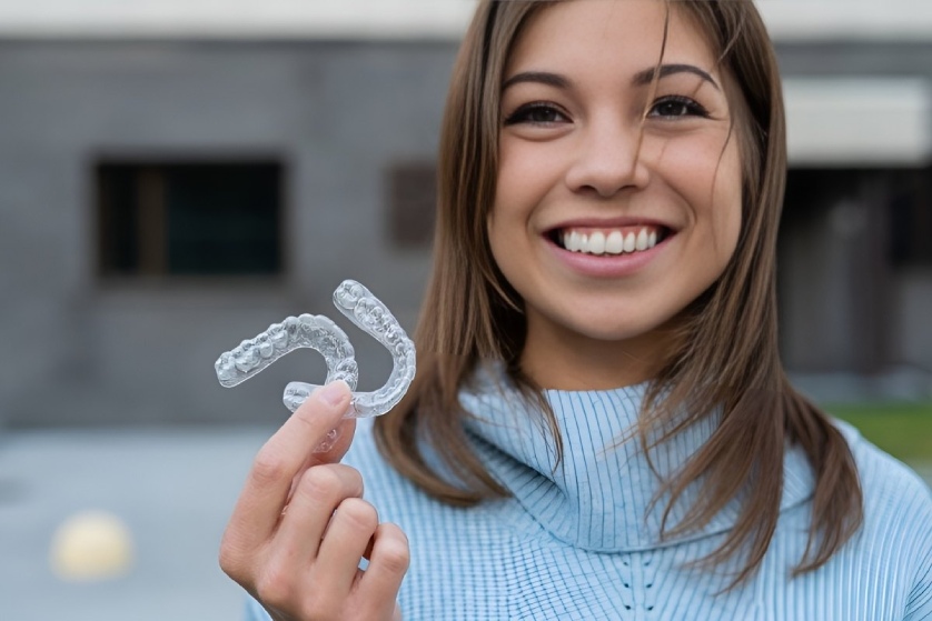
This information can support Invisalign treatment in Traverse City, MI Suttons Bay, MI Elk Rapids, MI and other orthodontic planning by helping us evaluate tooth positions, spacing, crowding, and bite relationships. Digital models can also make it easier to show patients what we are seeing and explain how treatment may change their smile.
Panoramic X-Ray
A panoramic X-ray provides a broad view of the upper and lower jaws, including erupted teeth, developing teeth, roots, and supporting structures.
This type of image can help us evaluate tooth development, identify missing or impacted teeth, and understand how permanent teeth are positioned beneath the gums. It is especially useful during growth and development when planning early orthodontic treatment.
Lateral Cephalogram Imaging
Lateral Cephalometric X-rays provide a side-view image of the head and help us evaluate relationships between the teeth, jaws, facial profile, and surrounding structures.
These measurements can be especially helpful when bite correction, growth, or jaw relationships are important parts of treatment planning. They give Dr. Lints another perspective when determining how orthodontic movement may affect both function and facial balance.
GETTING STARTED WITH INVISALIGN
If Invisalign sounds like it is the right fit for you, call or text us to set up a complimentary consultation. During this consultation, Dr. Lints and team will:
- 1. Review your dental and medical history
- 2. Perform a complete oral exam
- 3. Help you determine if Invisalign is the best route for you
- 4. Create you a customized treatment plan with before and after simulation
- 5. Discuss flexible payment plan options and insurance benefits if applicable
 231.922.7210
 Traverse City